Ankle range of motion.
Normal values for dorsiflexion, plantarflexion, inversion and eversion, how to line up a goniometer, and how to measure from a camera.
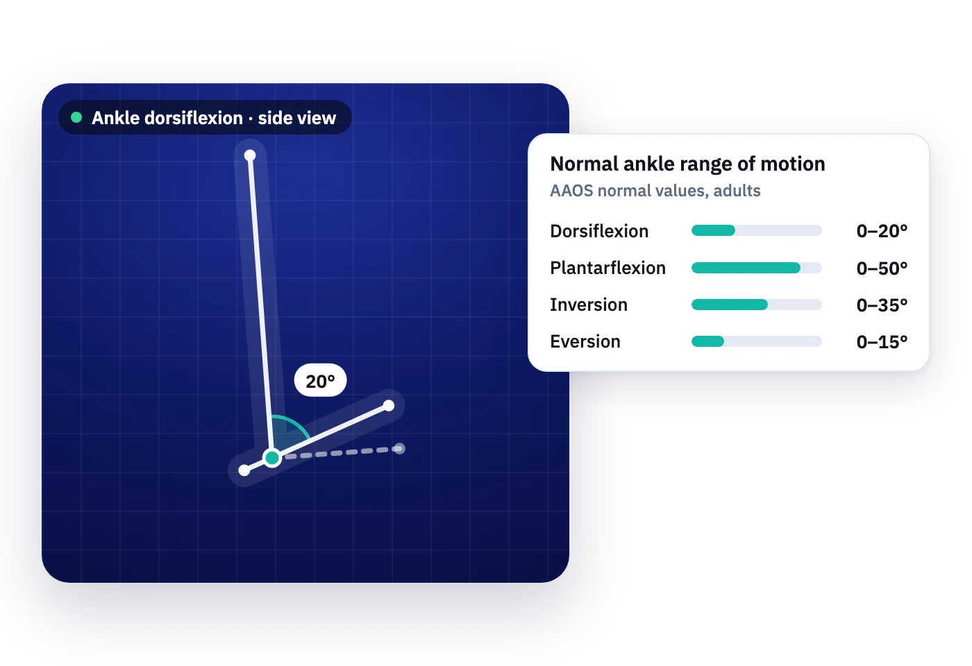
Ankle dorsiflexion drives squat depth, stair descent and gait, so a few degrees matter. The neutral position is the foot at 90 degrees to the leg, which means every reading starts from an offset.
Normal ankle range of motion
AAOS reference values, with the CDC average for healthy adults aged 20 to 44 where it was measured.
| Motion | AAOS | CDC average women / men |
|---|---|---|
| Dorsiflexion | 0–20° | 14° / 13° |
| Plantarflexion | 0–50° | 62° / 55° |
| Inversion | 0–35° | – |
| Eversion | 0–15° | – |
By age and sex
From the CDC Normal Joint Range of Motion Study: more than 600 healthy people measured by physical therapists with universal goniometers. Women had more range than men in nearly every joint, and range fell with age in both.
| Motion | Women 20–44 | Men 20–44 | Women 45–69 | Men 45–69 |
|---|---|---|---|---|
| Dorsiflexion | 13.8° | 12.7° | 11.6° | 11.9° |
| Plantarflexion | 62.1° | 54.6° | 56.5° | 49.4° |
How to measure ankle range of motion with a goniometer
Standard goniometric alignment. Knee position changes dorsiflexion a lot, so record whether the knee was bent or straight.
Dorsiflexion and plantarflexion
- Position
- Seated with the knee bent 90°, or supine with the knee straight (reads lower)
- Axis
- Lateral malleolus
- Stationary arm
- Lateral midline of the fibula, toward the fibular head
- Moving arm
- Parallel to the lateral aspect of the fifth metatarsal
- Watch for
- Toes extending, the foot inverting or everting, and a change in knee position
Inversion and eversion
- Position
- Seated with the knee bent 90°, ankle in neutral
- Axis
- Anterior aspect of the ankle, midway between the malleoli
- Stationary arm
- Anterior midline of the tibia, toward the tibial tuberosity
- Moving arm
- Anterior midline of the second metatarsal
- Watch for
- Hip rotation. Expect poor agreement between testers for these motions
Measuring ankle range of motion with a camera
Dorsiflexion and plantarflexion read best side-on with the camera lowered to ankle height. Weight-bearing tests such as the knee-to-wall lunge read well on camera, because the shin angle is easy to see.
- From a photo: the free online goniometer lets you tap the axis and both segments on a picture and read the angle. Nothing is uploaded.
- Live: MotionArc tracks the joints from any phone, tablet or laptop camera and measures the movement as it happens, in the clinic or on a video call.
Why two clinicians get different numbers
A goniometer reading depends on the person holding it. Finding the bony landmarks, lining up the arms and positioning the patient all add variation, and that variation is larger between two clinicians than for one clinician measuring twice.
- Small changes between two clinicians may not be real. At the shoulder, a true change measured by two different therapists needs about 15°, compared with 6° to 11° when the same therapist measures both times. Agreement between testers is weakest for shoulder extension and internal rotation.
- The instrument matters. At the knee, the smallest difference you can trust is about 6° with an inclinometer, 10° with a long-arm goniometer, 12° with a smartphone app and 14° with a short-arm goniometer or a visual estimate.
- Video can work. Shoulder angles measured over a video call with an on-screen protractor came within 0.2° to 5° of goniometer readings on average, and most smartphone apps studied show adequate reliability.
- What helps: a written protocol for patient position and landmarks, the same instrument each time, the same clinician where possible, and a note of how the measurement was taken.
Measuring from a camera takes out the hand-held step. Nobody has to line up the goniometer arms by hand, and the same view can be set up visit after visit.
Early access is limited, and we set up every account personally.
More reference values: normal range of motion chart for every joint. Other joints: Shoulder · Hip · Knee · Neck · Low back.
Sources
- American Academy of Orthopaedic Surgeons. Joint Motion: Method of Measuring and Recording. Chicago: AAOS; 1965.
- AAOS values as tabulated in a Jones & Bartlett Learning clinical handbook, chapter 1 (sample chapter). Link
- Hendriks et al. Plastic and Reconstructive Surgery Global Open. 2020. Supplement: normal range of motion values according to the AAOS. Link
- Soucie JM, Wang C, Forsyth A, et al. Range of motion measurements: reference values and a database for comparison studies. Haemophilia. 2011;17(3):500-507. Reference values from the CDC Normal Joint Range of Motion Study. Link
- Norkin CC, White DJ. Measurement of Joint Motion: A Guide to Goniometry. 5th ed. Philadelphia: F.A. Davis; 2016.
- University of West Alabama Athletic Training. Goniometry lab: joint measurement procedures. Link
- Baumbach SF, et al. 2014 (effect of knee position on ankle dorsiflexion). Link
- Elveru RA, Rothstein JM, Lamb RL. Goniometric reliability in a clinical setting: subtalar and ankle joint measurements. Phys Ther. 1988. Link
- Mullaney MJ, et al. Physiother Theory Pract. 2010;26:327 (reliability of shoulder range of motion measurement). Link
- Riddle DL, Rothstein JM, Lamb RL. Goniometric reliability in a clinical setting: shoulder measurements. Phys Ther. 1987;67:668. Link
- Hancock GE, et al. J Exp Orthop. 2018 (reliability of knee range of motion measurement methods). Link
- Sahu et al. J Shoulder Elbow Surg. 2022;31:201 (shoulder range of motion by video conference versus goniometer). Link
- Keogh JWL, et al. Reliability and validity of clinically accessible smartphone applications to measure joint range of motion: a systematic review. PLOS ONE. 2019;14:e0215806. Link
Measure ankle range of motion from any camera.
No goniometer, no wearables. The same movement, measured the same way, every visit.
Ankle range of motion questions
Straight answers. If yours is not here, request access and ask us anything.
About 20° by AAOS values, measured from the neutral position where the foot is at 90° to the leg. Healthy adults in a CDC study averaged about 13°.
Center the axis on the lateral malleolus, align the stationary arm with the fibula toward the fibular head and the moving arm parallel to the fifth metatarsal. Read the change from the 90° neutral position.
Both are useful, but they give different numbers. With the knee bent the gastrocnemius is slack, so the test reflects the soleus and joint. With the knee straight it includes gastrocnemius length and reads lower. Record which you used.
Yes. MotionArc measures range of motion, form and balance from any phone, tablet or laptop camera, in the clinic or live on a video call. Early access is open to a small group of professionals.
Clinicians sign a business associate agreement (BAA) with us before any real patient data is entered. The free goniometer on this site runs in your browser and never uploads photos.
Request early access
Tell us a bit about your work. We review every request personally and set you up ourselves.